Ventral Tegmental Area (VTA) of Tsai
The VTA was first characterized by Chiao Tsai (or Cai Qiao) in the opossum's brain in 1925. (Tsai went on to become a professor at Fudan University and later National Shanghai Medical College. He worked with Lord Edgar Adrian in Cambridge from 1930-32.)
From Tsai, Chiao. The optic tracts and centers of the opossum. Didelphis virginiana, Journal of Comparative Neurology, 39(1925)173
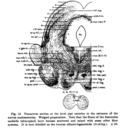
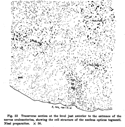
The cells contained in the nucleus opticus tegmenti are small with a few medium-sized fusiform ones in its dorsal part (fig. 23). Medial to this nucleus lies a group of cells, the nucleus tegmenti ventralis (nuc.teg.v.), which Kosaka and Hiraiwa (’15) and Castaldi (’23) regard as a part of the suhstantia nigra. The former authors have even gone so far as to assert that the fusiform cells in the nucleus opticus tegmenti probably belong to the substantia nigra too. However, according to our observations, the nucleus opticus tegmenti and the nucleus tegmenti ventralis are distinctly separated by a clear cell-free space lying between them, and the fusiform cells in these two nuclei are comparatively smaller than those in the substantia nigra. Furthermore, the nucleus tegmenti ventralis, being imbedded in the peduncular fibers of the corpus mamillare and the tracti mamillo- and olfacto-tegmentalis, seems to have functional relation with these bundles, that is, olfactory connection, thus differing from the non-specific character of the substantia nigra connections.

